Platelet-rich plasma (PRP) has diverse applications in dermatology, including wound healing and aesthetics. However, confusion persists regarding terminology, classification, and preparation methods. Inconsistencies in protocols and limited product characterization hamper standardization.
Moreover, expensive commercial Platelet-rich plasma kits limit wider usage. Addressing these issues can enhance the accessibility and effectiveness of plasma treatments in dermatology.
Plasmolifting World concentrates on creating various instruments and techniques using PRP tubes to speed up body cell and tissue repair and healing.
What is the process?
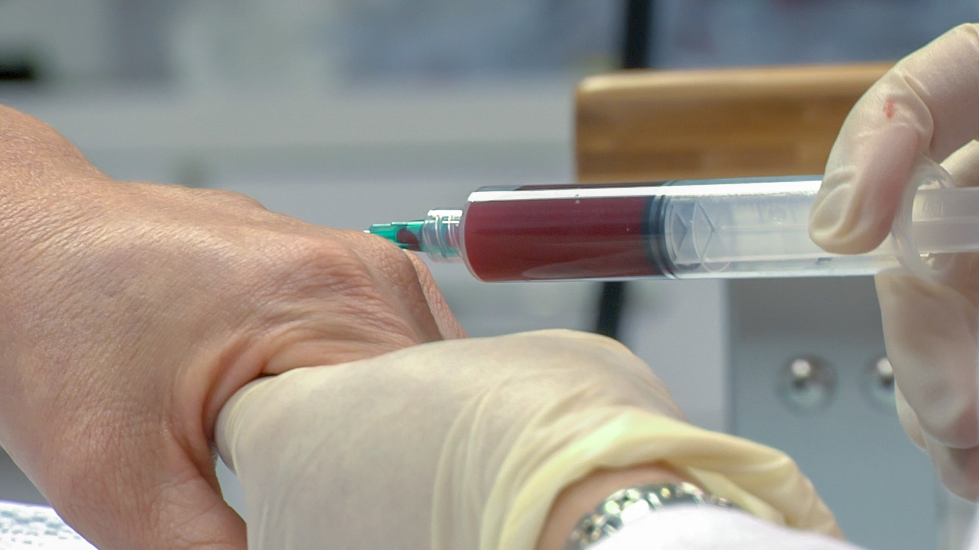
Platelets are crucial blood cells for wound healing, helping clot formation and supporting cell growth. For preparing plasma injection, blood is taken and centrifuged to separate components, including PRP, which is then extracted for medical use.
Research indicates injecting areas with high platelet concentrations can stimulate tissue growth and cellular healing. It is often combined with bone graft therapies for tissue repair and employed in treating various muscular, skeletal, or skin conditions by medical professionals.
Preparation of Platelet-rich plasma
Platelet-rich plasma is created using a patient’s blood sample drawn during the procedure. The volume of plasma generated typically ranges from 3-5 CC, depending on factors such as
- The patient’s initial platelet count
- The specific method employed
- The equipment utilized, following a 30 CC venous blood draw.
To prevent platelet activation before usage, an anticoagulant, such as citrate dextrose A, is added to the blood drawn during the blood draw. It makes use of a customized “tabletop cold centrifuge” machine. Costs associated with preparation are far lower than with commercial kits.
Principles of preparation
Platelet-rich plasma is typically prepared through a technique called differential centrifugation. This process adjusts the acceleration force to separate various cellular components based on their specific gravity.
There are two primary methods for its preparation:
- The PRP method
- The buffy-coat method.
In the first approach, whole blood with anticoagulants is initially collected in tubes. The first centrifugation step, performed at a constant acceleration, separates red blood cells (RBCs) from the remaining whole blood volume.
A second centrifugation step then concentrates platelets suspended in the smallest final plasma volume. This method yields plasma for various medical applications.
Three layers of the WB split after the first spin step.:
- A top layer primarily made up of platelets and WBC
- A thin, middle coating known as the buffy coat that is abundant in WBCs
- A bottom layer that is primarily made up of RBCs.
The upper layer and the superficial buffy coat are carefully transferred into an empty sterile tube to produce pure Platelet-rich plasma. In contrast, the entire buffy coat layer and a few remaining red blood cells (RBCs) are transferred to create leucocyte-rich Platelet-rich plasma.
Subsequently, a second centrifugation step is carried out with the centrifugal force adjusted to just enough to facilitate the formation of soft pellets, primarily composed of erythrocytes and platelets, at the tube’s bottom.
The upper portion, predominantly platelet-poor plasma (PPP), is removed. The resulting pellets are blended with the lower third (5 ml) plasma to generate Platelet-Rich Plasma.


